
Eye Exam
Clinical Basis
Diabetic retinopathy is a highly specific vascular complication of both type 1 and type 2 diabetes with prevalence strongly related to both the duration of diabetes and the level of glycemic control. It is the most frequent cause of new cases of blindness among adults aged 20 to 74 in developed countries. Glaucoma, cataracts and other disorders of the eye occur earlier and more frequently in people with diabetes.
Clinical Guidelines
- Optimize glycemic control to reduce the risk or slow the progression of diabetic retinopathy [A].
- Optimize blood pressure and serum lipid control to reduce the risk or slow the progression of diabetic retinopathy [A].
Screening Recommendations1
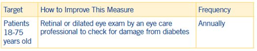
- Patients with type 2 diabetes should have an initial dilated and comprehensive eye examination by an ophthalmologist or optometrist at the time of the diabetes diagnosis [B].
- If there is no evidence of retinopathy for one or more annual eye exams, then exams every two years may be considered.
- If any level of diabetic retinopathy is present, subsequent dilated retinal examinations for patients with type 1 or type 2 diabetes should be repeated at least annually by an ophthalmologist or an optometrist.
- If retinopathy is progressing or sight-threatening, then examinations will be required more frequently [B].
Coding and Documentation Guidance
Refer patients to optometrist/ophthalmologist for dilated retinal exam. In addition to documenting the type of eye exam performed (including findings), submit from the following CPT II codes and HCPCS procedure codes based on report received from optometrist/ophthalmologist.
CPT II
- 2022F: Dilated eye exam with interpretation by an ophthalmologist or optometrist documented and reviewed with evidence of retinopathy (DM)
- 2023F: Dilated retinal eye exam with interpretation by an ophthalmologist or optometrist documented and reviewed; without evidence of retinopathy (DM)
- 2024F: 7 standard field stereoscopic retinal photos with interpretation by an ophthalmologist or optometrist documented and reviewed with evidence of retinopathy (DM)
- 2025F: 7 standard field stereoscopic retinal photos with interpretation by an ophthalmologist or optometrist documented and reviewed; without evidence of retinopathy (DM)
- 2026F: Eye imaging validated to match diagnosis from 7 standard field stereoscopic retinal photos results documented and reviewed with evidence of retinopathy (DM)
- 2033F: Eye imaging validated to match diagnosis from 7 standard field stereoscopic retinal photos results documented and reviewed; without evidence of retinopathy (DM)
- 3072F: Low risk for retinopathy (no evidence of retinopathy in the prior year)
1 American Diabetes Association. 12. Older adults: Standards of Medical Care in Diabetes 2019. Diabetes Care 2019;42 (Suppl. 1):S139–S147
